
Figure 7. DCA treatment decreased caspase-3 activation in the mouse brain after HI.
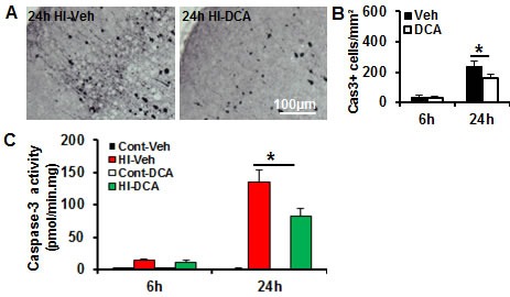
A. Representative pictures show staining for the active form of caspase-3 in the brain cortex at 24 h after HI. B. Quantification of the caspase-3-positive cells in the IHC in the cortex. C. The caspase-3 activity (as indicated by DEVD cleavage) was assayed in the brain cortical homogenate at 6 h and 24 h after HI in the DCA-treated mice (n = 7) and the vehicle-treated mice (n = 8). *p < 0.05.