
Figure 3. MiR-708 inhibits renal cancer cell proliferation and promotes apoptosis.
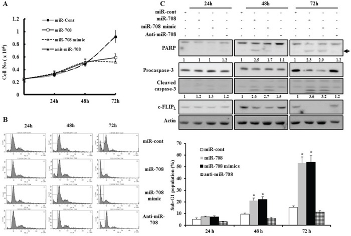
A. Caki cell proliferation analyzed using trypan blue dye exclusion assays 24 h, 48 h, and 72 h after transfection with miR-708, miR-708 mimics, anti-miR-708, or miR-cont. Cell proliferation was significantly inhibited after transfection with miR-708 or miR-708 mimics compared to cells transfected with miR-cont. B. Caki cells were transfected with miR-708, miR-708 mimics, anti-miR-708, or miR-cont for 24 h, 48 h, or 72 h. Apoptosis was analyzed as a sub-G1 fraction by FACS (Histogram, left panel). Flow cytometry analysis of apoptotic cells (Graph, right panel). The data is reported as the mean ± SD (n = 3). * indicates P < 0.05 versus miR-cont-transfected cells. C. Transfection with miR-708 or miR-708 mimics induced cleavage of PARP and procaspase-3 after 48 h. Equal amounts of cell lysates (40 μg) were subjected to electrophoresis and analyzed by Western blotting with c-FLIPL, PARP, and caspase-3 antibodies. Proteolytic cleavage of PARP is indicated by the arrow. Actin was used as a loading control in all immunoblots. The relative levels of cleaved PARP and caspase-3 in miR-transfected cells were expressed as the ratio of the densitometric values for each protein to those of actin.