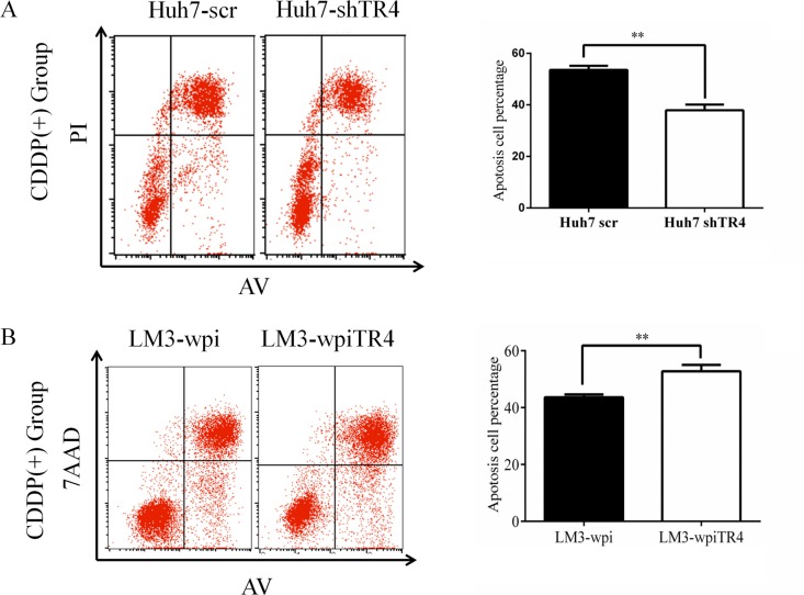
Figure 5. Higher TR4 expression resulting in a higher cell apoptosis in HCC cells treated with cisplatin.
(A) Huh7-scramble/shTR4 cells were treated with 6 μg/ml cisplatin for 48 h. Apoptosis was assessed as described in Materials. Quantification is shown on the right. Comparison among groups was performed using Student's test. (B) LM3-vector/TR4 cells were treated with 10 μg/ml cisplatin for 48 h. Flow cytometry (left) and comparison among groups (right) are shown as in (A). All assays were were performed in triplicate (*P < 0.05, **P < 0.01, ***P < 0.001).

