
Figure 7. The P-gp activity.
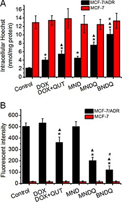
(A) and expression (B) of MCF-7/ADR and MCF-7 cells after treatment with free DOX, (DOX + QUT), MND, MNDQ and BNDQ. In the experiment of P-gp activity, the cells were incubated for 4 h in the drug-free medium (control), free DOX, (DOX + QUT), MND, MNDQ or BNDQ, and Hoechst 33342 (4 μM) were added to the culture media and incubated for another 0.5 h. Then the cells were washed, lysed and analyzed fluorimetrically for the intracellular content of the dye. The dye concentration was standardized by the protein concentration which was measured with BCA assay. In the experiment of P-gp expression, the cells were cultured with the drug-containing media or drug-free media (control group) for 48 h, and then the cells were trypsinized, collected, and resuspended. PE-conjugated mouse anti-human monoclonal antibody against P-gp was use to label above cells, and the fluorescent intensities of the cells were determined. Measurements were performed in triplicate and data are presented as mean ± SD (n = 3). (*p < 0.01, compared to control group; ▴p < 0.01, compared to DOX group; #p < 0.01, compared to MNDQ group).