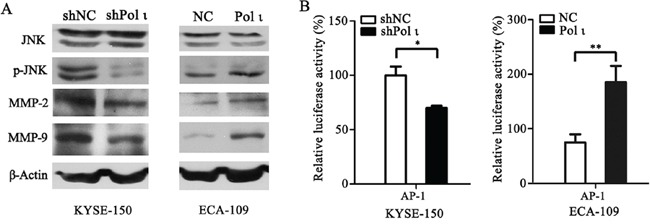
Figure 6. Pol ι enhances MMP-2 and MMP-9 expression through activating the JNK-AP-1 pathway.

A. western blot analysis of the expression levels of JNK, p-JNK, MMP-2 and MMP-9 in Pol ι overexpression and knockdown cell lines.β-actin levels served as loading control. B. cells were seeded in 24-well plates and transfected with pAP1-Luc vector for 36h. The luciferase activity was analyzed by a dual-luciferase reporter system. The luciferase activity was normalized by co-transfection with 50 ng pRL-SV40 vector and analysis of Ranilla activity (*p < 0.05, **p<0.01, Student's t test).
