
Figure 1. The GRK2/CXCR2/AKT pathway is changed in B lymphoma cells infected by KSHV.
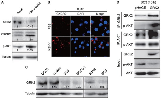
A. Western blotting analysis of GRK2, CXCR2 and phosphorylated AKT in KSHV latently infected BJAB cells (KSHV-BJAB) and uninfected control cells (BJAB). Numbers labeled under the bands were the relative intensities of the bands following calibration for loading by house-keeping proteins. Some of the following Western blotting figures have similar densitometry analysis. B. IFA for the expression of CXCR2 (red) in BJAB cells infected with KSHV (KSHV; bottom) or treated with PBS (PBS; top). DAPI (blue) stains nuclei (Original magnifications, x1000). C. Western blotting analysis of GRK2 in KSHV-infected PEL cells (BC3 and BCBL-1 cells) and KSHV-negative lymphoma cells (DG75, Loukes, and BJAB cells), respectively. D. Co-immunoprecipitation of GRK2 and AKT in GRK2-overexpressing BC3. BC3 cells were transduced with lentivirus-GRK2 (GRK2) or its control (pHAGE) for 48 h, respectively, and then subjected to immunoprecipitation with antibody against GRK2 (IP: GRK2) or AKT (IP: AKT). At the same time, cell lysates were immunoblotted with indicated antibodies (Input).