
Figure 1. Induction of MCT4 and TRAP1 and inhibition of SDH in Early Preneoplastic Foci (EPF) and preneoplastic nodules.
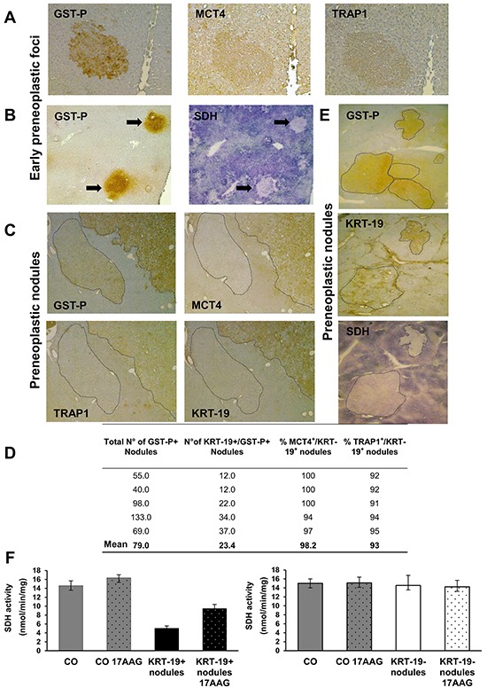
A. IHC on serial sections of livers from rats sacrificed 4 weeks after DENA (1 week after PH) shows staining for both MCT4 and TRAP1 in a GST-P+ EPF (x10). B. Histochemical reaction showing impaired SDH activity in GST-P+ EPF (arrows) (x4). C. IHC on serial sections of livers from rats sacrificed 10 weeks after DENA shows MCT4 and TRAP1 staining in a GST-P+/KRT-19+ nodule. Absence of MCT4 and TRAP1 staining is observed in an adjacent GST-P+/KRT-19− nodule (dotted area, x4). D. Percentage and number of KRT-19+/MCT4+ and KRT-19+/TRAP1+ nodules with respect to the total amount of KRT-19+ nodules. E. Photomicrographs showing a perfect match between GST-P/KRT-19 induction and SDH activity inhibition in nodules (dotted areas) (x1.25). F. SQR enzymatic activity of SDH in control livers (CO) and GST-P+/KRT-19+ (left) or GST-P+/KRT-19− (right) nodules; when indicated, 17-AAG was added 5 min before starting recordings.