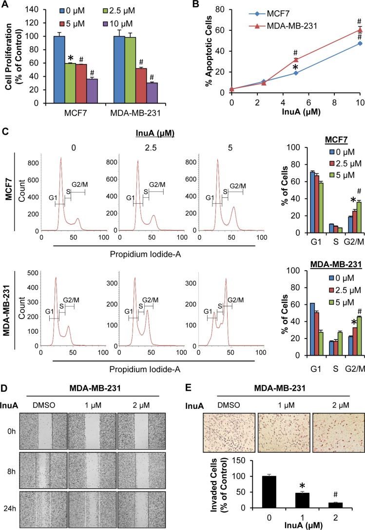
Figure 2. In vitro anti-breast cancer activity of InuA.
(A) Antiproliferative effects of InuA. MCF7 and MDA-MB-231 cells were exposed to various concentrations (0, 2.5, 5, and 10 μM) of InuA for 24 h, followed by measurement of cell proliferation via the BrdUrd assay. The proliferative index is in comparison to untreated cells; (B) Induction of apoptosis by InuA. MCF7 and MDA-MB-231 cells were treated with InuA (0, 2.5, 5, and 10 μM) for 48 h, followed by measurement of apoptosis using Annexin V assay/flow cytometry; (C) Effects of InuA on cell cycle progression. MCF7 and MDA-MB-231 cells were treated with InuA (0, 2.5 and 5 μM) for 24 h, followed by determination of cell cycle distribution using flow cytometry; (D&E) Effects of InuA on metastasis. MDA-MB-231 cells were treated with InuA (0, 1, and 2 μM) for 24 h, the migration ability was measured by a wound healing assay (D); and cell invasion ability was measured using a Transwell cell invasion assay (E). All assays were performed in triplicate and repeated three times. (*P < 0.05 and #P < 0.01) DMSO, dimethyl sulfoxide.

