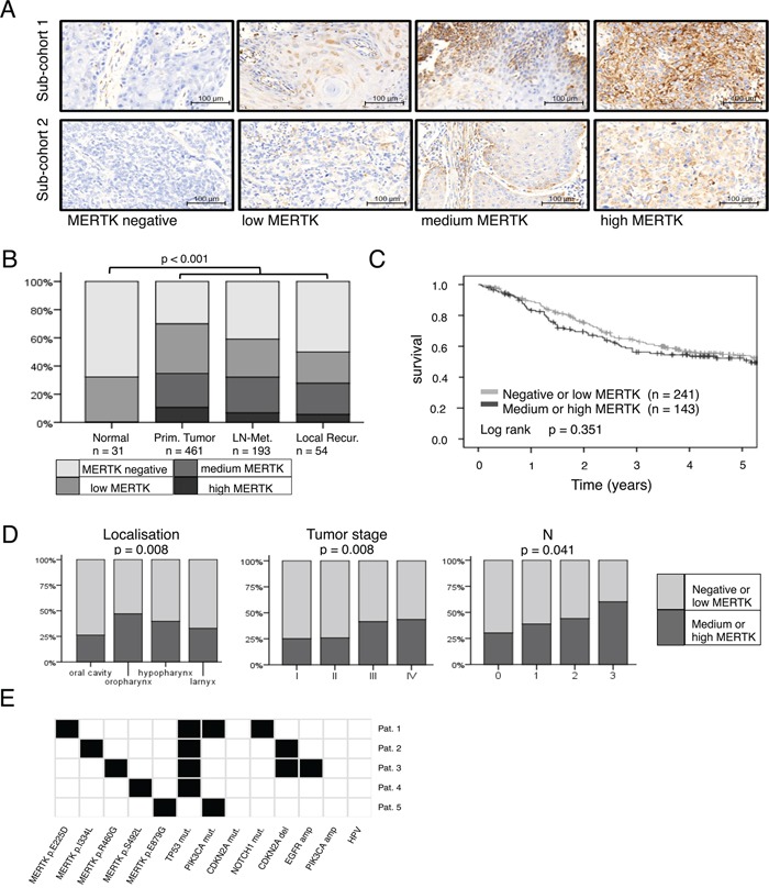
Figure 1. MERTK expression is increased in head and neck cancer.

A. Representative cores from primary tumors for each MERTK staining intensity for both staining protocols/sub-cohorts. B. Protein expression of MERTK in normal mucosa (n = 31), primary tumors (n = 461), lymph node metastases (n = 193) and local recurrences (n = 54). C. Kaplan-Meier estimates for overall survival of patients with negative or low MERTK protein expression compared to patients with medium or high expression. D. MERTK protein expression in different primary tumor localizations, tumor- and N-stages. E. MERTK mutations in HNSCC (n = 5/279) and additional mutations found in these five patients. (B: Fisher test: Monte Carlo, 100 000 random samples; C: log-rank test; D: Fisher test: Monte Carlo, 100 000 random samples).
