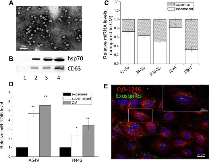
Figure 2. Extracellular miR-1246 exists in non-exosomes associated form.
(A) Exosomes released from A549 cells were observed under an electron microscopy. (B) Hsp70 and CD63 protein expressions in the exosomes purified from the medium with depleted exosomes FBS (lane 1) and the CM of A549 cells with a number of 2.5 × 10⁶ (lane 2), 5 × 10⁶ (lane 3), and 1 × 10⁷ (lane 4). (C) Comparison the levels of five miRNAs in the exosome-free supernatant and exosome-enriched pellet of the CM from A549 cells. The total expression level of each miRNA was set to 1. (D) Relative miR-1246 levels in the exosomes, exosome-free supernatant and the CM from A549 and H446 cells. The miR-1246 level in exosomes was set as control. Data were presented as means ± SEMs of five independent experiments (*P < 0.05,** P < 0.01). (E) Localization of exosomes and extracellular miR-1246 in the recipient A549 cells. The CM was collected from A549 cells labeled with DiO and Cy3-labeled miR-1246 and was then used to incubate other recipient A549 cells for 24 h. The recipient A549 cells with red fluorescence Cy3-labeled miR-1246, green fluorescence exosomes, and DAPI stained nuclear with blue fluorescence were fixed and observed under a confocal microscopy.

