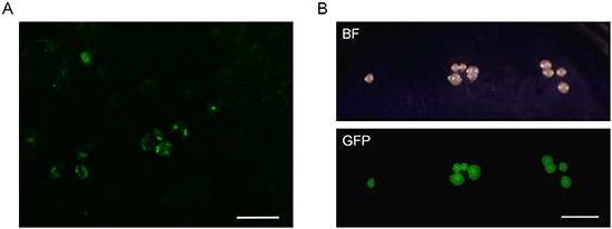
Figure 2. Imaging tumor-targeting Salmonella typhimurium A1-R in the FDCS PDOX.

A. FDCS was resected from a PDOX model after 4 weeks treatment of S. typhimurium A1-R and a subsequent two weeks without treatment. FV1000 confocal microscopy. Scale bar = 100 μm. B. Colonies of S. typhimurium A1-R isolated from the tumor of the bacterially-treated FDCS PDOX after 4 weeks treatment and a subsequent 2 weeks without treatment.
