
Figure 2.
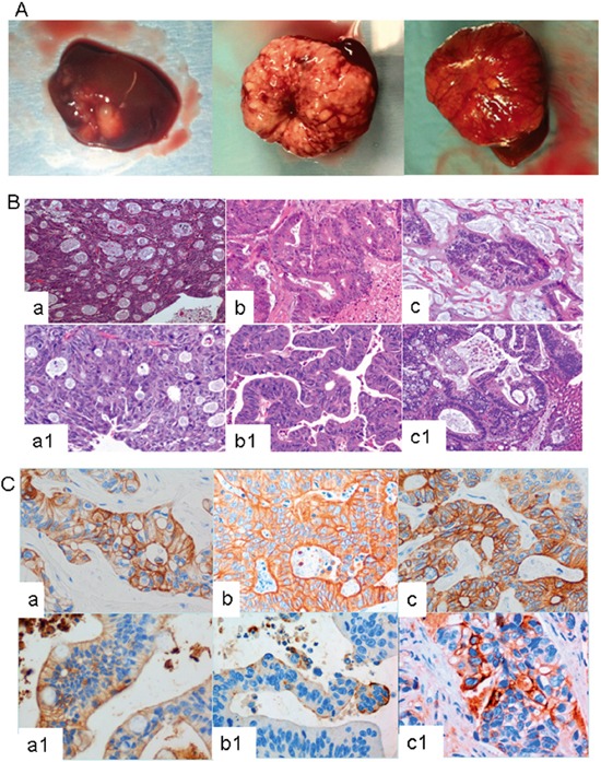
A. Three representative examples of CRC liver metastasis growing orthotopically in murine liver. B. and C. Human CRC liver metastasis and their derived PDX lines show similar pathological features. B) Hematoxylin and eosin staining and C) cytokeratin 20 staining of CRC human liver metastasis and their derived PDX lines. In these figures, a/b/c correspond to the human tumors, and a1/b1/c1, to the PDX derived tumors.