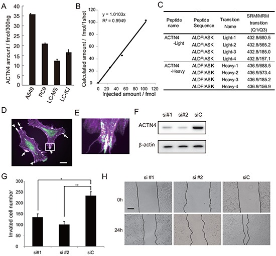
Figure 2. Effect of ACTN4 knockdown using siRNA of ACTN4 on in vitro cell migration ability.

A. Absolute amount of ACTN4 protein in non-small cell lung cancer (NSCLC) cell lines by multiple reaction monitoring (MRM) using mass spectrometry (MS). B. Standard curve of MRM with ACTN4 peptide. C. Peptide sequence and MRM transition for actinin-4 quantitation using MS. Bold letters indicate amino acid residues labeled with stable isotope (13C and 15N). D, E. Immunocytochemical analysis of ACTN4 (green) and phalloidin (violet) staining of A549 cells. Arrows indicate co-stained lamellipodia, and arrowheads indicate co-stained filopodia. Bar in D indicates 10 μm. F. Western blot analysis of ACTN4 expression in A549 cells transfected with ACTN4 (si#1 or si#2) or control (siC) siRNA. G. Boyden chamber assay of the effect of ACTN4 siRNAs on cell invasion. *P < 0.05, **P < 0.01 (t-test). H. Representative photographs of wound healing by cells transfected with siRNA of ACTN4 or control siRNA. The cells are shown at wounding (0 h) and 24 hours later (24 h). Bar indicates 100 μm.
