
Figure 5. Effect of ACTN4 knockdown using shRNA of ACTN4 on cell metastatic ability in an in vivo animal model.
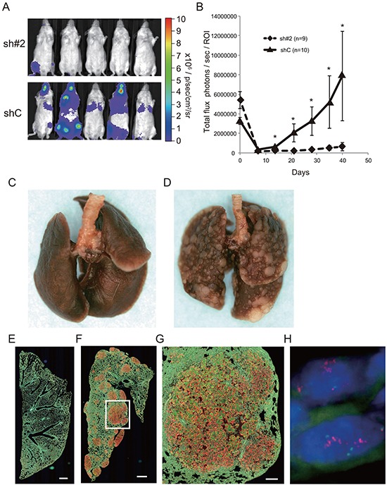
A, B. Evaluation of luciferase signals from the injected mice using an in vivo imaging system (IVIS). (A) Representative photographs of luciferase signals from mice injected with the different A549 cells (40 days after injection). (B) Alterations in luciferase signal intensities over time after cell injection (diamonds; sh#2, and triangles shC. Bars; SEM, and *P < 0.01 Mann Whitney t-test). C, D. Representative murine lungs on day 40 (C, sh#2; D, shC). E–G. Immunohistochemical analysis of murine lung on day 40 with anti-ACTN4 (red) and anti-cytokeratin 19 (green) antibodies (E, sh#2; F, G; shC). Protein overexpression of ACTN4 was detected in the metastatic lesions of murine lung. Bar in E and F indicates 500 μm, and that in G indicates 100 μm. H. Representative fluorescence in situ hybridization analysis of ACTN4 in the metastatic region of animal models.