
Figure 2. Effects of PIM kinase inhibition on mTORC1 downstream targets in GBM cells.
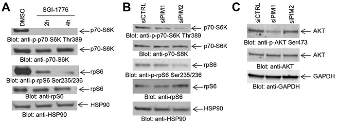
A. LN229 cells were treated with SGI-1776 (5 μM) for the indicated times. Equal amounts of total cell lysates were subjected to SDS-PAGE followed by immunoblotting with antibodies against the phosphorylated forms of p70-S6K (pThr389), rpS6 (pSer235/236), or against HSP90 as indicated. Equal amounts of cell lysates from the same experiment were analyzed in parallel by SDS-PAGE and immunoblotted with antibodies against p70-S6K or rpS6, as indicated. B. LN229 cells were transfected as indicated with control siRNA or siRNAs directed against PIM1 or PIM2. Equal amounts of total cell lysates were subjected to SDS-PAGE followed by immunoblotting with antibodies against the phosphorylated forms of p70-S6K (pThr389), rpS6 (pSer235/236), or against HSP90 as indicated. Equal amounts of cell lysates from the same experiment were analyzed in parallel by SDS-PAGE and immunoblotted with antibodies against p70-S6K or rpS6, as indicated. C. LN229 cells were transfected with control siRNA and siRNAs directed against PIM1 or PIM2. Equal amounts of total cell lysates were subjected to SDS-PAGE followed by immunoblotting and membranes were simultaneously incubated with antibodies directed against the phosphorylated form of AKT (pSer473), AKT or GAPDH, followed by visualization in a ChemiDoc MP Imaging System (BioRad) as described in Materials and Methods.