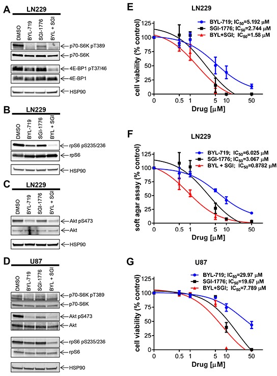
Figure 3. Effects of simultaneous inhibition of PIM and PI3K on cell viability and transformation of GBM cells.

A. LN229 cells were treated with SGI-1776 (5 μM) and with BYL-719 (10 μM) for 90 min. Lysates were analyzed by SDS-PAGE and immunoblotted with the indicated antibodies for HSP90 and the phosphorylated forms of p70-S6K and 4E-BP1. Subsequently, membranes were stripped and reprobed with antibodies against p70-S6K and 4E-BP1, as indicated. B. Same experiment as in A using antibodies for HSP90 and the phosphorylated form of rpS6 followed by stripping and reprobing with antibodies against rpS6 as indicated. C. LN229 cells were treated and processed as in A. Membranes were incubated simultaneously with antibodies against HSP90, the phosphorylated form of AKT on Ser473 and total AKT, followed by visualization in a ChemiDoc MP Imaging System (BioRad) as described in Materials and Methods. D. U87 cells were treated and processed as in A. Proteins were immunoblotted with antibodies for p70-S6K, rpS6 and their phosphorylated forms (pThr389 and pSer235/236, respectively) and HSP90 simultaneously. The membrane was stripped and reprobed with antibodies the phosphorylated form of AKT on Ser473 and total AKT simultaneously. E. LN229 cells were plated in 96-well plates and treated with increasing concentrations of the PIM inhibitor SGI-1776 and/or PI3K inhibitor BYL-719 for 5 days. Cell viability was assessed using WST-1 proliferation assay. Results represent the means ± SEM of 3 independent experiments, each done in triplicates. F. LN229 cells were plated in 96-well plates in soft agar and treated with increasing concentrations of PIM inhibitor SGI-1776 and PI3K inhibitor BYL-719 for 7 days. Colony formation was quantified using the fluorescent cell stain CyQUANT GR Dye (Cell Biolabs Inc.) in the Synergy HT Plate reader. Data are expressed as percentages of control DMSO treated samples. Results represent the means ± SEM of 3 independent experiments, each done in triplicates. G. Same as E using U87 cells. Cell viability was assessed using WST-1 proliferation assay. Results represent the means ± SEM of 3 independent experiments.
