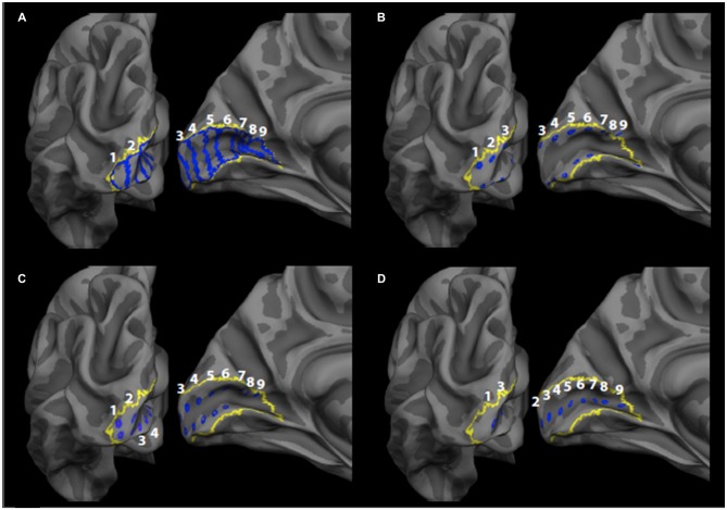
Figure 1.
Primary visual cortex (V1) regions of interest (ROIs). The label boundaries (blue) for the (A) bar ROIs, (B) gyrus crown ROIs, (C) sulcus wall ROIs, and (D) sulcus depth ROIs are shown within the Freesurfer V1 label boundary (yellow) on the fsaverage brain. ROIs are numbered 1–9, such that one corresponds to the most posterior ROI (most central representation) and nine corresponds to the most anterior ROI (most peripheral representation).

