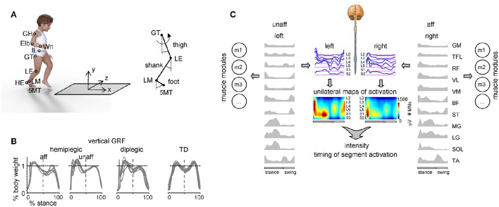
Figure 1.
Experimental setup and recorded spatiotemporal gait parameters. (A) Schematic view of the experimental setup and recorded body landmarks and angles. Elb, elbow; GH, gleno-humeral joint; HE, heel; IL, ilium; LE, lateral femur epicondyle; LM, lateral malleolus; Wri, wrist. (B) Examples of vertical ground reaction forces in one representative child with hemiplegia (9.9 years), one diplegia (6.1 years) and one TD (11.8 years) child. The data from several strides were superimposed. Since children typically made more than one foot placement on the force plate (so that we could not isolate the force of one leg throughout the whole stance phase), the ground reaction forces in the first and second half of the stance were taken from different strides (and separated by the vertical dashed line). (C) Example of the spatiotemporal patterns of muscle and motoneuron activity along the rostrocaudal axis of the spinal cord during walking in one child with hemiplegic. Recorded EMG waveforms were used to reconstruct muscle modules (mi) and output pattern of each segment of each side of the lumbosacral enlargement (blue curves). Same pattern is plotted in a color scale (right calibration bar) using a filled contour plot (middle). Patterns are plotted vs. the normalized gait cycle.

