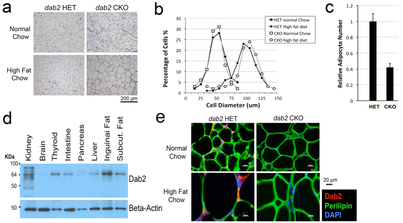
Figure 2. Impact of high fat diet on cell size and number of Dab2-deficient mice.
(a) H&E sections of gonadal adipose tissues are shown for dab2 CKO and HET controls fed either normal or high fat chow. (b) The images were analyzed using the MetaMorph Image Analysis Software for the diameter of the adipocytes as an indication of cell size. The cell size distribution is shown as percentage of cells at each increment of diameter. HFD induced a similar increase of adipocyte cell size in both dab2 CKO and HET mice. (c) The relative number of adipocytes in gonadal adipose tissues from dab2 CKO and HET mice on HFD was estimated based on the tissue mass and average cell diameter. (d) Dab2 protein expression was determined by Western blot in protein extracts from various tissues dissected from the mice. It was confirmed that Dab2 proteins were completely absent in all tissues from the dab2 CKO mice. (e) Analysis by immunofluorescence microscopy of Dab2 and perilipin-1 in gonadal fat tissues from dab2 CKO and HET mice fed with either normal or high fat diet.

