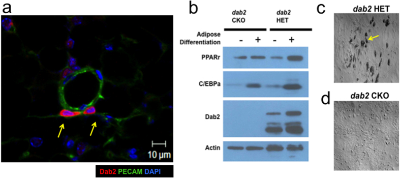
Figure 6. Adipose differentiation from cells of the stromal vascular fractions.
Dab2-positive cells were identified to locate around blood vessels in adipose tissues. Cells of the stromal vascular fractions were isolated from inguinal adipose tissues of two-month-old dab2 HET and CKO male mice. The cells were cultured for 1 week and then were subjected to differentiation for 2 days. (a) Inguinal adipose was sectioned and stained for PECAM (green, a marker for vesicular endothelial cells), Dab2 (red), and DAPI (blue). Dab2-positive cells (indicated by arrows) were found located around blood vessels in the adipose tissues. (b) Lysates were prepared from pre- and post-differentiation, and were analyzed by Western blot. All gels were derived from the same cell lysates and processed in an identical condition. (c) The morphology of the dab2 HET cells are shown. The arrow indicates the presence of lipid droplets contained in pre-adipocytes of HET (but rare in CKO) cells.

