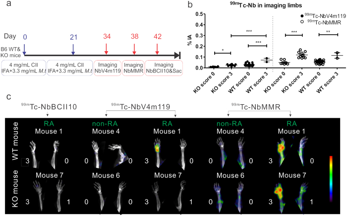
Figure 1. SPECT/CT quantification and imaging of 99mTc-NbV4m119, 99mTc-NbMMR or 99mTc-NbBCII10 accumulation in WT and CRIg−/− (KO) C57BL/6J mice with CIA after the onset of the arthritis symptoms.
(a) Experimental timelines for C57BL/6J mice with CIA; IFA = Incomplete Freund’s Adjuvant; M.t = Mycobacterium tuberculosis strain H37RA; Sac = mice were sacrificed. SPECT/micro-CT imaging was performed 3 h post intravenous injection of 99mTc-labeled NbV4m119, NbMMR or NbBCII10. (b) Signals obtained in the joints with score 3 and joints with score 0 were grouped in four symptomatic and four asymptomatic mice (two WT and two KO mice in each group). Data are expressed as mean values of % of injected activity (%IA) of 99mTc-Nanobodies obtained from the limbs (front paws, metatarsal joints and ankles). **p < 0.01 and ***p < 0.001. (c) Representative SPECT/μCT image of front paws in CIA mice, 3 h post injection with 99mTc-labeled NbV4m119, NbMMR or NbBCII10. Clinical scores are indicated next to each joint. Representative images of 1 mouse per group are shown using National Institutes of Health color scale and are scaled to maximum in the whole image. The data are representative of two independent experiments.

