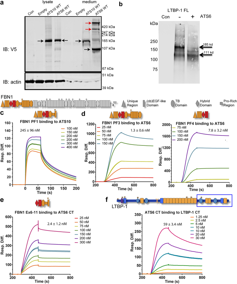
Figure 6. ADAMTS6 but not ADAMTS10 is processed and catalytically active, binds microfibrillar proteins.
(a) Western blot against V5 tag of ADAMTS10 (ATS10 WT) and ADAMTS6 (ATS6 WT) overexpressed proteins using lentivirus. The SDS-PAGE was run under reducing conditions. Both full-length molecules expressed well in this system. Unprocessed ADAMTS10 was present in medium and cell lysates, whereas ADAMTS6 was processed in medium (black arrows). Some larger bands may be aggregated forms of the processed molecule (red arrow), which are possibly trimeric and tetrameric in nature. (b) Full-length LTBP-1 was treated with full-length recombinant ADAMTS6 overnight at 37 °C (molar ratio LTBP-1:ADAMTS6 3:1). Western blot analysis using anti-LTBP-1 C-terminal antibody showed a reduction in full-length LTBP-1 with a relative molecular mass of 185 kDa, and an increase in intensity of a 111 kDa degradation product. The cleavage site of BMP-144, indicated by ‘X’ on LTBP-1 domain map (Fig. 6F), also generates a 110 kDa C-terminal fragment. Control lane (con) contains ADAMTS6 only. (c–f) Biacore sensorgrams showing that: (c) Full-length ADAMTS10 binds the fibrillin-1 PF1 fragment, and (d) full-length ADAMTS6 binds to recombinant N-terminal fibrillin-1 (PF3 and PF4). (e) To further resolve the binding regions, it was found that ADAMTS6 CT regions (Fig. 4) bound fibrillin-1 Ex6–11 fragment16. The binding of C-terminal ADAMTS6 was mapped to exons 6–8 of FBN1, indicated in red. (f) C-terminal ADAMTS6 binds to LTBP-1 C-terminal region (indicated by box). Binding was analysed using Surface Plasmon Resonance, and the response difference (Resp. Diff.) for each experiment was plotted against time (s). Resp. Diff. is the ligand-immobilized flow cell minus the control flow cell.

