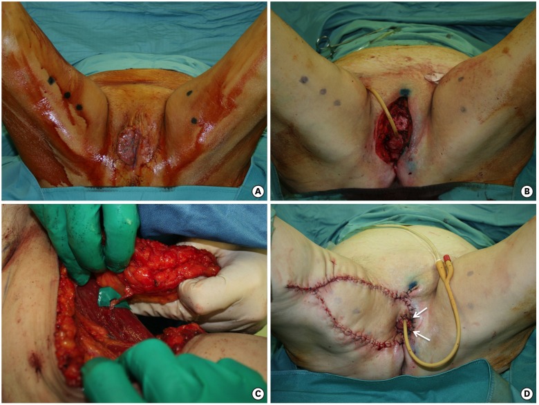
Fig. 2.
(A) Tumor on the right hemivulva. (B) Radical vulvectomy, wider on the right side. (C) The flap harvested on the medial surface of the right thigh, pedicled on a medial circumflex femoral artery perforator. (D) The flap advanced in a V-Y fashion. The white arrows indicate the part of the flap that goes beyond the midline to surround the urethra and vagina.

