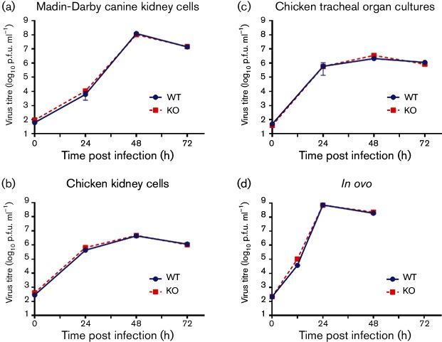
Fig. 2.
In vitro growth curves of UDL01 WT and KO viruses. Cellular substrates were inoculated at a low m.o.i. with either WT (blue circles) or KO (red squares) UDL01 influenza virus. The level of infectious virus was determined at times specified. Data represent the means of at least three independent experiments±sd. (a) MDCK cells and (b) primary cKCs were inoculated at an m.o.i. of 0.0001. (c) Chicken tracheal organ cultures were inoculated with 100 p.f.u. (d) The 10-day old Rhode Island Red chicken eggs were inoculated into the allantoic cavity with 100 p.f.u.

