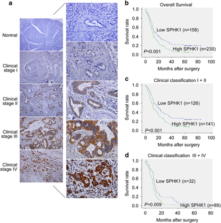
Figure 5.
SPHK1 expression in pancreatic cancer and normal pancreatic tissues via immunohistochemistry. (a) IHC staining indicated that SPHK1 expression is upregulated in human pancreatic cancer tissues (clinical stages I–IV) compared with normal pancreatic tissues (left panel: magnification × 100; right panel: magnification × 400). (b) Kaplan–Meier curves of pancreatic cancer patients with low versus high expression levels of SPHK1 (n=388; P<0.001). (c, d) Significance of the difference between the curves of SPHK1 high-expressing and low-expressing patients was compared in clinical stages I−II (c) and clinical stages III−IV (d) patient subgroups. P-values were calculated based on the log-rank test.

