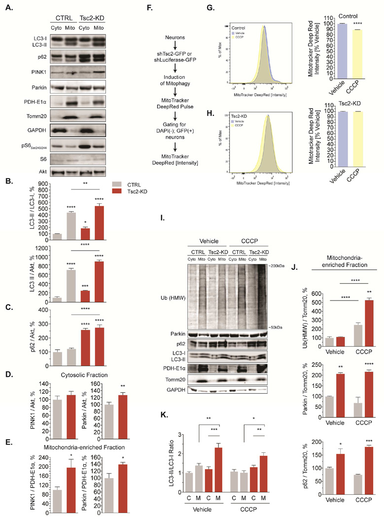
Figure 5. Global mitophagic flux is impaired in Tsc2-deficient neurons.
(A-E) Representative western blots and quantification of several proteins involved in mitophagy in cytosolic and mitochondria-enriched fractions from cortical neurons (DIV7/8, n=12 experiments)
(F-H) Mitophagic flux induced by CCCP (1 µM, 24hr) quantified using flow cytometry in cortical neurons (DIV11, n=6 × 10⁶ recorded events from 6 experiments).
(I-K) Western blotting for several proteins involved in mitophagy in cytosolic and mitochondria-enriched fractions from cortical neurons (DIV7/8) treated with CCCP (1 µM, 24hr) or vehicle (n= 3–6 experiments).
C, Cyto=cytosolic fraction, HMW=high molecular weight, M, Mito=mitochondria-enriched fraction, mono=monomeric; *p<0.05, **p<0.01, ***p<0.001, ****<p.0001. See also Figure S5.

