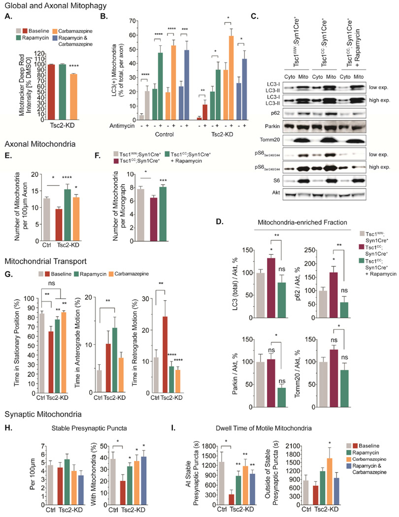
Figure 7. The impact of mTOR-dependent and mTOR-independent stimulation of autophagy on mitochondrial dynamics, turnover and function.
(A) Quantification of mitophagic flux using flow cytometry in cortical neurons (DIV11) treated with rapamycin (20nM, 24hr) or carbamazepine (100 µM, 24hr, n=3×10⁶ recorded events per condition from 3–6 experiments).
(B) Recruitment of Venus-LC3 labeled autophagosomes to axonal mitochondria following acute Δ Ψm-depolarization with antimycin A (40 µM) in hippocampal neurons (DIV7/8) pretreated with rapamycin, carbamazepine or a combination of both drugs (n>35 axons per condition from 5–10 experiments).
(C&D) Western blotting for several proteins involved in mitophagy in cytosolic and mitochondria-enriched fractions from cortex of Tsc1cc;Syn1Cre+ mice (n=8), Tsc1CC;Syn1Cre+ mice treated with rapamycin (n=4), and Tsc1ww;Syn1Cre+ littermates (n=8, PND 21).
(E) Number of mitochondria per 100 µm axon in hippocampal neurons (DIV 7/8) pretreated with rapamycin or carbamazepine (n>20 axons per condition from >4 experiments).
(F) Number of mitochondria in callosal projection axons from Tsc1cc;Syn1Cre+ mice (n=2), Tsc1cc;Syn1Cre+ mice treated with rapamycin (n=3), and Tsc1ww;Syn1Cre+ littermate controls (n=3, PND 21) using transmission electron microscopy.
(G) Mitochondrial transport in the mid axon of hippocampal neurons (DIV7/8) pretreated with rapamycin or carbamazepine (n>15 axons per condition from 3–5 experiments).
(H) Number of stable presynaptic puncta per 100 µm axon and the percentage of stable presynaptic puncta supported by mitochondria in hippocampal neurons (DIV7/8) pretreated with rapamycin (20nM, 24hr), carbamazepine (100 µM, 24hr) or a combination of both drugs (n=14 axons per condition from 7 experiments).
(I) Dwell time of motile mitochondria at and outside of stable presynaptic puncta in hippocampal neurons (DIV7/8) pretreated with rapamycin, carbamazepine or a combination of both drugs (n=14 axons per condition from 7 experiments).
Cyto=cytosolic fraction, Mito=mitochondrial fraction, ns=not significant; *p<0.05, **p<0.01, ***p<0.001, ****<p.0001. See also Figure S6.

