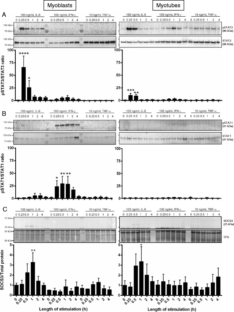
Fig. 1.

IL-6 and IFN-γ stimulation initiate JAK/STAT signaling in C2C12 myoblasts and myotubes in vitro. C2C12 proliferating myoblasts (left) or differentiated day 5 myotubes (right) either remained unstimulated (0) or were incubated with 100 ng/mL rmIL-6, 100 ng/mL rmIFN-γ, or 10 ng/mL rmTNF-α for 0.25, 0.5, 1, 2, or 4 h at 37 °C + 5 % CO2. At the conclusion of stimulation, cells were lyzed and subject to SDS-PAGE and western immunoblotting for either pSTAT3/STAT3 (a), pSTAT1/STAT1 (b), or SOCS3 and total protein (TPS; c). Representative immunoblots are shown for each probe with n = 1 per time-point. Three individual experiments were performed, and bands were quantified to produce the graphs shown. Data are expressed as mean ± SEM. Statistical analysis was performed using a one-way ANOVA with a Fisher’s LSD post hoc multiple comparisons test to determine the effect of treatment. *P < 0.05, **P < 0.01, ***P < 0.001, ****P < 0.0001 compared to unstimulated cells
