
Fig. 2.
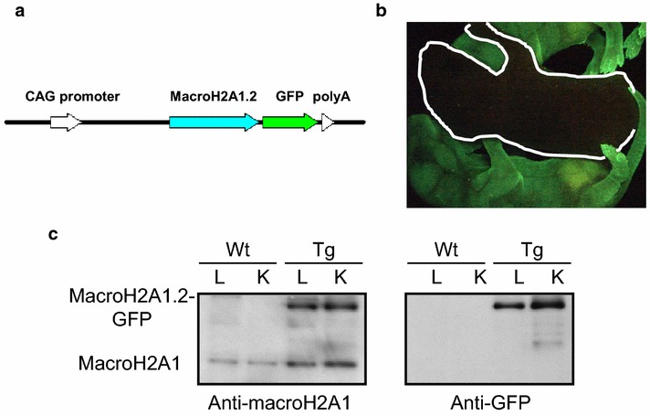
MacroH2A1.2-GFP transgenic mice (adapted from [26], licence no. 3766141157990). a Structure of the vector expressing macroH2A1.2–EGFP (pCXMH2A/EGFP). Mouse macroH2A1.2 tagged with EGFP was expressed under the control of the CAG promoter with a cytomegalovirus early enhancer element and a chicken b-actin promoter. b Stereomicroscopic analysis. The macroH2A1.2–EGFP transgenic mouse (Tg) shows green fluorescence in the skin. The white line marks the position of a wild-type mouse. c Immunoblot analysis. Nuclear extracts (10 µg/lane) of liver (L) and kidney (K) were prepared from wild-type (WT) and transgenic (Tg) female mice and analyzed using anti-GFP and anti-macroH2A1 antibodies