
Fig. 7.
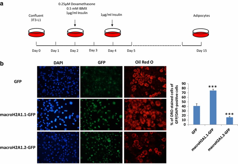
a Schematic protocol for 3T3-L1 differentiation from pre-adipocytes into mature adipocytes [28]. b Left panels 3T3-L1 pre-adipocytes with lentiviral-mediates stable expression of GFP, macroH2A1.1-GFP and macroH2A1.2-GFP were induced to differentiate into mature adipocytes as in (a). At the 15th day of differentiation, cells grown on coverslips were stained with Oil Red O (ORO) solution and counterstained with DAPI for nuclei. ORO was visualized with fluorescence microscopy. Right panel the percentage of ORO-stained cells was quantified over the total of GFP/DAPI-positive cells, averaging 10 different random fields in 3 separate experiments. Results are represented as mean ± S.E.M. ***p < 0.001 change versus GFP