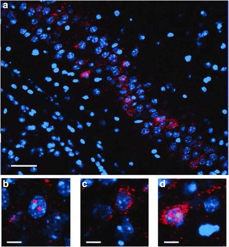
Fig. 2.

Representative images for Arc catFISH data in area CA3. a Fluorescence image showing Arc expression following exploration of identical (AA) environments (image taken at 20× 1 z stack). Scale bar: 50 μm. Example of neurons with foci only (b), cytoplasmic only (c) or cytoplasmic and foci Arc expression (d) (Scale bar: 100 μm). The Arc mRNA is illustrated in red and cell nuclei are indicated in blue (DAPI)
