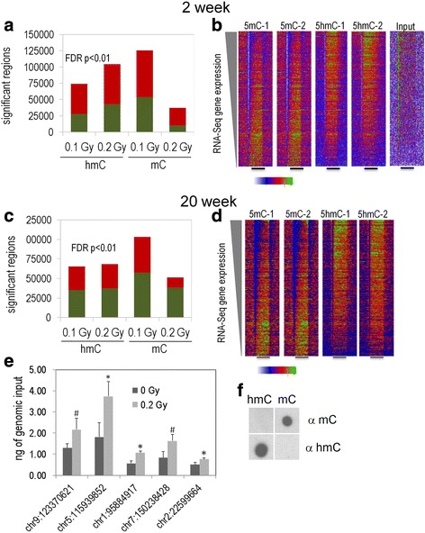
Fig. 5.

Number of genetic regions with significantly increased (green) and decreased (red) 5hmC and 5 mC at the 2- (a) and 20-week (c) time points. At both time points, there were more regions with up- and downregulated 5mC at the 0.1 than 0.2 Gy. Dip density histograms illustrating 5mC and 5hmC signal at RefSeq genes sorted by RNA-Seq gene expression levels at the 2-week (b) and 20-week (d) time points. e Real-time PCR validation of significantly upregulated DIP-Seq regions at the 0.2 Gy 2wk time point (samples selected based on ranked p-value). Real-time PCR was conducted with genomic input standard curves and 4 DIP biological replicates. *denotes p < 0.05 and #denotes p < 0.1. f The antibodies used to pull down 5mC and 5hmC regions do not cross react
