
FIGURE 2.
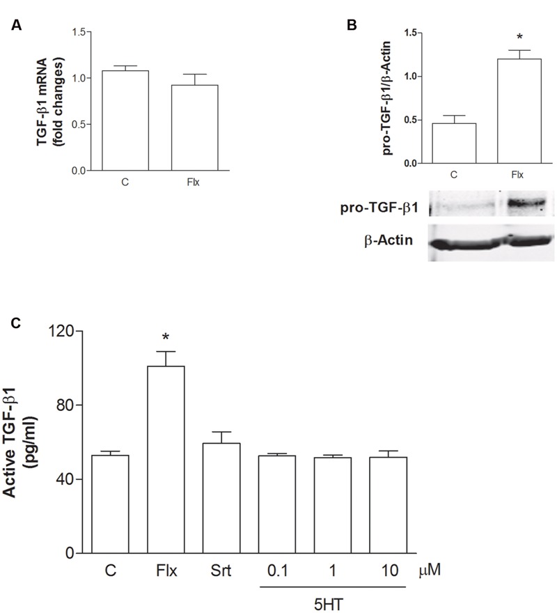
Fluoxetine increases the expression of pro-TGF-β1 and the release of active TGF-β1 from cortical astrocytes. (A) TGF-β1 mRNA levels obtained by Real-time RT-PCR in cultured astrocytes transiently exposed to fluoxetine (Flx; 1 μM) for 6 h are shown. Values were normalized by endogenous GAPDH mRNA levels and are represented as means + SEM for n = 4 for two independent experiments. (B) Representative immunoblots of pro-TGF-β1 (about 55 kDa) in total protein extracts from rat cortical astrocytes exposed to fluoxetine (Flx; 1 μM) for 24 h. Bars refer to the means ± SEM of the densitometric values of pro-TGF-β1 bands normalized against β-actin. Each experiment was repeated four times; ∗p < 0.05 vs. control by Student’s t-test. (C) Levels of active TGF-β1 in the medium of cultured astrocytes exposed for 24 h to fluoxetine (Flx; 1 μM), sertraline (Srt; 1 μM) or to increasing concentrations of serotonin (100 nM–10 μM) are shown. Values are means ± SEM of nine determinations; ∗p < 0.05 (Student’s t-test) versus untreated control astrocytes.