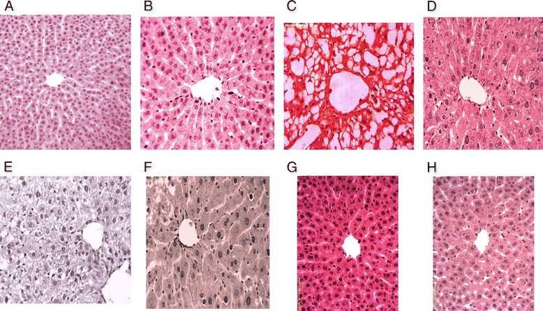
Fig. 3.

Histopathological studies of liver. Hematoxylin and eosin stain; 40× (a) Untreated control showing normal histoarchitecture of the hepatic tissues (b) vehicle treated (DMSO + olive oil) showing normal histoarchitecture of the hepatic tissues (c) CCl4 treated showing macrosteatosis (d) CCl4 + silymarin (100 mg/kg) treated showing almost normal histoarchitecture (e) CCl4 + FXM (200 mg/kg) treated showing microsteatosis (f) CCl4 + FXM (400 mg/kg) treated showing mild microsteatosis (g) FXM (200 mg/kg) treated showing normal histoarchitecture of the hepatic tissues (h) FXM (400 mg/kg) treated showing normal histoarchitecture of the hepatic tissues of rat. FXM; F. xanthoxyloides leaves methanol extract
