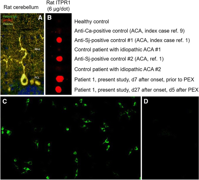
Fig. 1.

ITPR1-IgG as detected by a immunohistochemistry, b a dot-blot assay and c, d a cell-based assay (exemplary findings). a Binding of patient serum IgG and of a commercial antibody to ITPR1 to formalin-fixed mouse (as well as rat and primate, not shown) cerebellum tissue sections (MoL molecular layer, PCL purkinje cell layer). Binding of patient IgG is depicted in green (goat anti-human IgG Alexa Fluor® 488); binding of a commercial rabbit anti-ITPR1 antibody in red (goat anti-rabbit IgG Alexa Fluor® 568); yellow indicates binding of both antibodies. Cell nuclei were stained by 4',6-diamidino-2-phenylindole (blue colour). b Binding of IgG from the patient’s serum and from two previously reported ITPR1-IgG-positive patients [1] to purified native ITPR1 from rat brain in a dot-blot assay; by contrast, sera from four healthy and disease controls did not bind to ITPR1. A commercial goat anti-human IgG antibody labelled with IRdye®700 (Rockland, Limerick, PA) was used to detect bound patient IgG. c Binding of patient IgG to cells transfected with mouse ITPR1 (left) but not to mock-transfected HEK293 cells used as control substrate (right). Binding of patient IgG was visualized using a goat anti-human IgG secondary antibody labelled with fluorescein isothiocyanate. See the “Methods” section for details. ACA autoimmune cerebellar ataxia, PEX plasma exchange
