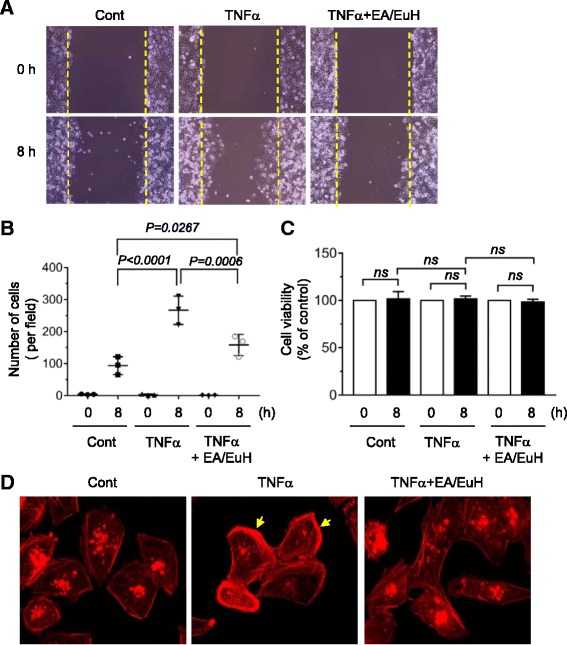
Fig. 2.

Effect of ethyl acetate fraction of E. humifusa Willd (EA/EuH) on TNFα-induced motility of MDA-MB-231 cells. a Cell migration assay. MDA-MB-231 cells were treated with or without EA/EuH (5 μg/mL) for 30 min, followed by exposure to TNFα (10 ng/mL). After 8 h, the representative field images were captured by an EVOS FL Auto Cell Imaging System. Dotted lines indicate the scraped boundaries at the beginning of the experiment. b Cells migrated into the gap area were quantified in a field of view using ImageJ software. The data shown represent the mean ± SD (n = 3). P value was analyzed by Sidak’s test. c Cell viability assay. MDA-MB-231 cells were treated with or without EA/EuH (5 μg/mL) for 30 min, followed by addition of TNFα (10 ng/mL). After 8 h, viable cells were determined using a Cell Counting Kit-8 (CCK-8). The data shown represent the mean ± SD. P value was analyzed by Sidak’s test. ns, not-significant. d MDA-MB-231 cells were treated with or without EA/EuH (5 μg/mL) for 30 min, followed by exposure to TNFα (10 ng/mL). After 12 h, the cells were stained with rhodamine-phalloidin (1:100) for 1 h and actin rearrangement was analyzed. Arrows indicate polarized F-actin
