
Figure 1.
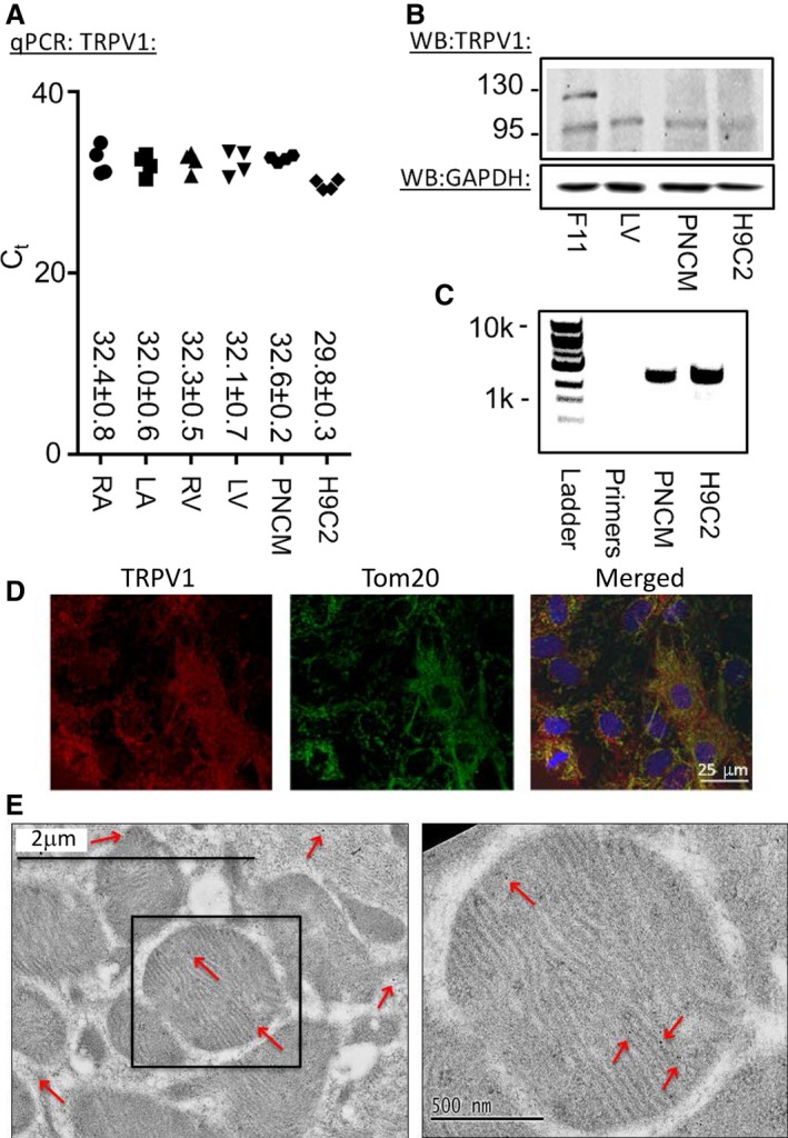
Biochemical evidence of TRPV1 is present in cardiomyocytes. A, qPCR for the 4 heart chambers (LV, LA, RV and RA), PNCMs, and the H9C2 cell line (n=4 biological replicates, each measured in triplicate). B, WB of total LV heart homogenate, PNCMs, and H9C2 cells. A dorsal root ganglion–derived cell line, F11, was used as a positive control. TRPV1 was intracellular (95 kDa) and glycosylated at the plasma membrane (120 kDa). GAPDH was used as a loading control (representative of 3 biological replicates). C, Reverse transcriptase PCR of PNCMs and H9C2 cells. D, Confocal imaging of TRPV1 in PNCMs with colocalization to TOM20, a specific mitochondrial membrane protein. E, Electron microscopy of mitochondria labeled for TRPV1 with immunogold particles (red arrows). LA indicates left atria; LV, left ventricle; PNCM, primary neonatal cardiomyocyte; qPCR, quantitative polymerase chain reaction; RA, right atria; RV, right ventricle; TRPV1, transient receptor potential vanilloid 1; WB, Western blot.