Figure 6.

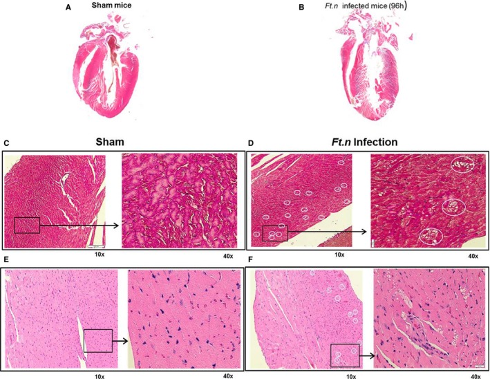
Presence of cardiac microlesions in Ft.n‐infected mice. Cross‐sections of hearts obtained from BALB/c mice after 96 hours of Ft.n challenge. (A) Four‐chamber view of hearts isolated from uninfected mice and (B) Ft.n‐infected mice. Overall size of the heart was reduced and showed significant tissue damage. Representative of 3 infected and uninfected hearts. The 4‐chamber view sections from uninfected (C) and Ft.n‐infected (D) mice were stained with Masson's trichrome to identify fibrotic replacement of cardiac tissue. Hematoxylin and eosin staining of uninfected (E) and Ft.n‐infected (F) heart sections and white circles show the regions of lesions by 10× magnification, and the area in the square was enlarged to 40× magnification. Cardiac microlesions were randomly dispersed throughout the septa and myocardium (N=6). White circled areas indicate lesion areas in the section.
