Figure 9.

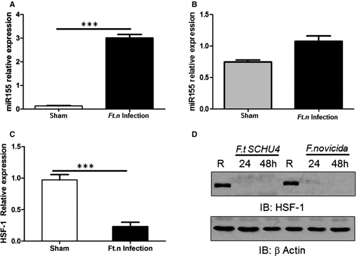
Ft.n infection enhances microRNA (miR)‐155 expression in the heart. Total RNA from hearts of Ft.n‐infected (25 CFUs) or uninfected BALB/c mice (3 per group) was extracted after 96 hours. Animals were sacrificed, hearts harvested immediately, cut into pieces to remove blood, washed 3×, and homogenized in RNA later. (A) miR‐155 expression was measured using the TaqMan MicroRNA assay kit and subsequent quantitative reverse‐transcriptase polymerase chain reaction (qRT‐PCR). N=2. (B) Myocytes from Ft.n‐infected or uninfected mice were isolated and lysed in TRIzol reagent, and total RNA was extracted. miR‐155 expression was determined by using the Taqman MicroRNA assay kit and subsequent qRT‐PCR. (C) Total RNA was extracted from infected and uninfected heart tissue to measure heat shock factor 1 (HSF‐1) expression by qRT‐PCR. N=3. (D) Bone‐marrow–derived macrophages were infected with Ft.n (MOI, 10:1) or left uninfected (R), and cells were lysed 24 and 48 HPI. Protein‐matched cell lysates were used to examine HSF‐1 levels by western blot and reprobed with anti‐β‐actin antibody as a loading control. Representative graph from 3 experiments (N=3; ***P<0.0005). CFU indicates colony forming unit; HPI, hours postinfection; IB, immunoblot; MOI, multiplicity of infection.
