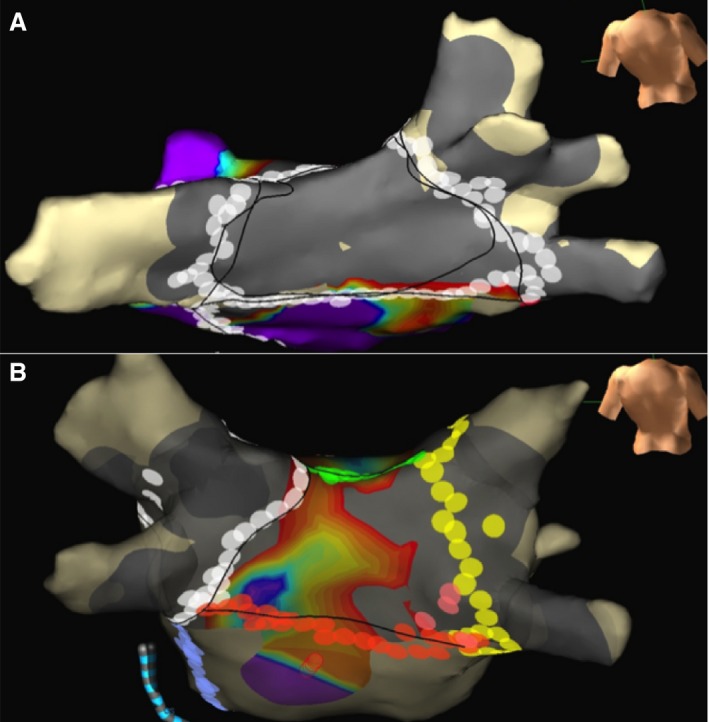
Figure 1.

A, Lesion sets to an isolated posterior wall, with a silent posterior wall on voltage mapping after completion of ablation. B, A patient unable to isolate the posterior wall despite lesion sets for posterior wall isolation, with electrical activity in areas of the posterior wall. Gray areas show bipolar peak‐to‐peak voltage during sinus rhythm <0.1 mV, and purple areas show bipolar voltage during sinus rhythm of >0.5 mV.
