Figure 4.

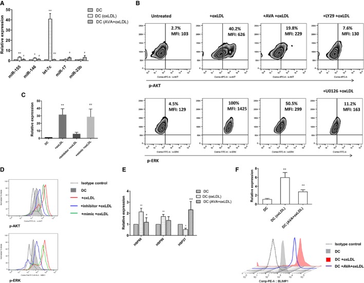
MicroRNA (miRNA) let‐7c and PI3k/ERK signaling pathways were involved in the effect of oxidized low‐density lipoprotein (oxLDL) and statins. A, Expression of miRNAs in dendritic cells (DCs) was tested by quantitative reverse transcription polymerase chain reaction (qRT‐PCR). The value was normalized as fold change to that of nontreated DC samples. Results represent the mean±SEM of 7 experiments. *P<0.05, **P<0.01, DC+oxLDL vs DC; # P<0.05, ## P<0.01, DC+ atorvastatin (AVA)+oxLDL vs DC+oxLDL. B, Phosphorylation of Akt and ERK induced in oxLDL‐treated DCs were confined by AVA. OxLDL‐induced phosphorylation of Akt and ERK were tested by phospho‐flow using specific antibody anti‐Akt (pS473) and anti‐MEK1 (pS298). Results show 1 representative experiment of 5. LY294002 (PI3k inhibitor) and U0126 (MEK inhibitor) are used as control. C, Inhibition of let‐7c in DCs was tested by qRT‐PCR. DCs at day 6 were treated with 10 μg/mL oxLDL for 24 hours. For inhibition of let‐7c, inhibitor of miRNA let‐7c, mimic control was transfected with Lipofectamine® RNAiMAX (Invitrogen) in a 20 nmol/L concentration at day 6. oxLDL was added 6 hours after transfection, and cells were further incubated for 24 hours. The value was normalized as fold change to that of nontreated DC samples. Results represent the mean±SD of 4 experiments. **P<0.01, DC+oxLDL vs DC; ## P<0.01, DC+inhibitor+oxLDL vs DC+mimic+oxLDL. D, Downregulation of let‐7c restricted the phosphorylation of Akt and ERK in the oxLDL‐treated DC. One of 4 experiments is shown. E, Expression of heat shock proteins (HSPs) in DCs was tested by qRT‐PCR. Results represent the mean±SD of 4 experiments. *P<0.05, **P<0.01, DC+oxLDL vs DC; # P<0.05, ## P<0.01, DC+AVA+oxLDL vs DC+oxLDL. F, Expression of B lymphocyte–induced maturation protein‐1 (BLIMP1) in DCs was tested by qRT‐PCR showing the mean±SD of 4 experiments, and flow cytometry showing a representative of 4 experiments. **P<0.01, DC+oxLDL vs DC; ## P<0.01, DC+AVA+oxLDL vs DC+oxLDL. MFI indicates median fluorescence intensity.
