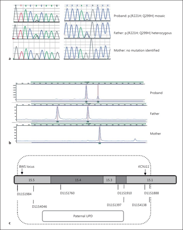
Fig. 1.
a Sequence electropherograms showing the p.R221H (c.662G>A; left panel) and p.Q299H (c.879G>C; right panel) KCNJ11 mutation(s). The proband is mosaic for both mutations, whilst the father is heterozygous. Neither mutation was found in the sample from the mother. b Electropherograms demonstrating the results of microsatellite analysis of the informative marker (D11S1984) in the proband (pancreatic tissue) and her parents (leukocytes). Data for the additional 6 informative markers are not shown. The x-axis indicates the product size [base pairs (bp)], and the y-axis the product quantity (arbitrary units). The results illustrate mosaic UPD with a larger peak for the paternal allele (180 bp) compared with the maternal allele (184 bp). c Results of chromosome 11 microsatellite analysis for DNA extracted from the proband (pancreatic tissue) and her parents (leukocytes). The 7 informative markers, which demonstrated paternal UPD, are shown along with their approximate location on chromosome 11. The position of KCNJ11 and the differentially methylated BWS locus are provided.

