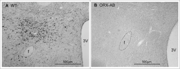
Figure 5.

Immunohistochemical demonstration of orexin-contained neurons in coronal sections through the caudal hypothalamus from a wild-type (WT) rat (A) and from an ORX-AB rat (B). In the WT animal, but not in the ORX-AB animal, there were orexin-positive neuronal cell bodies in the perifornical region. f, Fornix; 3V, third ventricle.
