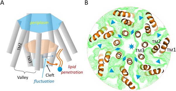
Figure 4.

Putative mechanism of adaptation of MscS. A: Schematics of lipid penetration. B: Channel connections within the Ec‐MscS complex. TM helices (orange ribbons) are obtained from the crystal structure of the open form (PDB ID: 2VV5). The complex is viewed from the cytosolic side, with cytosolic domains removed for clarity. Channels within the TM complex were calculated with probes of 1.4 Å radius and illustrated with green meshes. The central pore, clefts between TM3a helices, and valleys between TM1‐TM2 hairpins are marked with a star, diamond symbols, and triangle symbols, respectively.
