Figure 6.

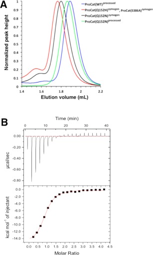
Zymogen and processed ProCat are structurally and functionally distinct. (A) Size exclusion analysis of the ProCat constructs. Plot of the elution volume and the normalized UV absorption at 280 nm for ProCat(WT)processed(blue), ProCat(Q152H)zymogen(red), ProCat(Q152N)zymogen(black), and ProCat(Q152N)processed(green). ProCat(S386A)processed eluted at the same location as ProCat(Q152H)zymogen and has been left out of this figure for clarity. (B) ITC measurements showing the background‐subtracted heats per injection (top panel) and binding isotherms (bottom panel) for the titration of WT‐ProCat PCSK9 with a peptide mimetic of the EGF‐A domain of the LDLr . Integrated heats were fit by non‐linear regression to a model for a single set of sites. The three zymogen constructs did not show binding to peptide (data not shown).
