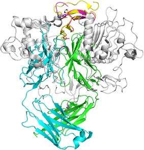
Figure 4.

Comparison of the EGFR (1MOX) and LY3016859 binding sites. Structures are aligned by TGFα. TGFα from 1MOX is shown in yellow, and LY3016859 bound TGFα is shown in magenta. EGFR is shown in grey while the LY3016859 Fab is colored cyan (LC) and green (HC). A Cd2+ ion is observed in the 1MOX structure (yellow sphere) and is bound by TGFα similarly to the Zn2+ ion in the LY3016859 bound structure. It is clear from the superposition of the complexes that EGFR and LY3016859 cannot simultaneously bind TGFα, and this provides a structural basis for the neutralization of TGFα by LY3016859.
