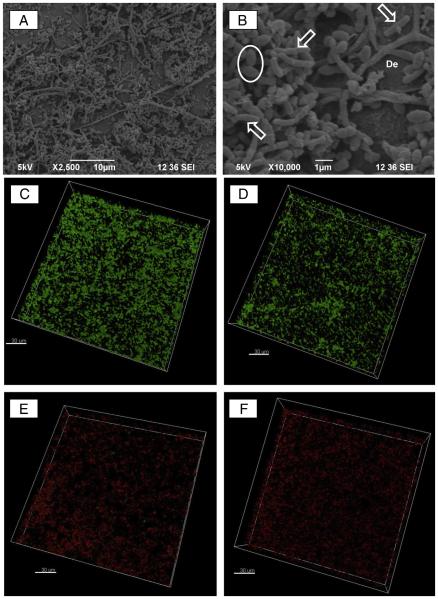
Figure 1.
Representative scanning electron microscopic micrographs of a 7-day biofilm formed by dual (A. naeslundii and E. faecalis) bacterial species. (A)A lower-magnification image showing a homogeneous distribution of the 2 bacterial cells; (B) a higher-magnification image of A revealing the rod-shaped A. naeslundii and cocci-shaped E. faecalis bacterial cells over the dentin (De) surface. CLSM images were collected in the sequential illumination mode by using 488-nm and 552-nm laser lines. Live bacteria presenting intact cell membranes were dyed green (SYTO 9), whereas dead bacteria with damaged membranes were stained red (propidium iodide). Fluorescent emission was collected in 2 HyD spectral detectors with a filter range set up to 500–550 nm and 590–655 nm for green (SYTO 9) and red dye (propidium iodide), respectively. CLSM images of (C) 7-day dual-species biofilm (negative control) growth inside dentinal tubules, (D) dual-species biofilm exposed to antibiotic-free nanofibers, (E) triple antibiotic-containing nanofibers, and (F) TAP solution for 7 days (bar = 30 μm).

