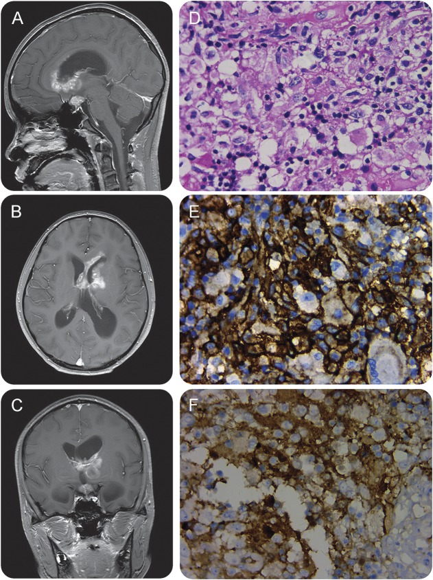
Figure. MRIs and histopathology of the biopsy specimens.
Sagittal (A), axial (B), and coronal (C) gadolinium-enhanced T1-weighted MRIs demonstrate extensive enhancing extra-axial masses in the bilateral ependymal/subependymal zones and suprasellar region, with perilesional edema and severe obstructive hydrocephalus. Hematoxylin & eosin–stained (D) specimen shows a dense proliferation of mixed inflammatory cells. Small lymphocytes predominate, but scattered large histiocytes and plasma cells are also observed. Histiocytic cells showing CD68 (E) and S-100 (F) immunoreactivity. Magnification 400× (D–F).

