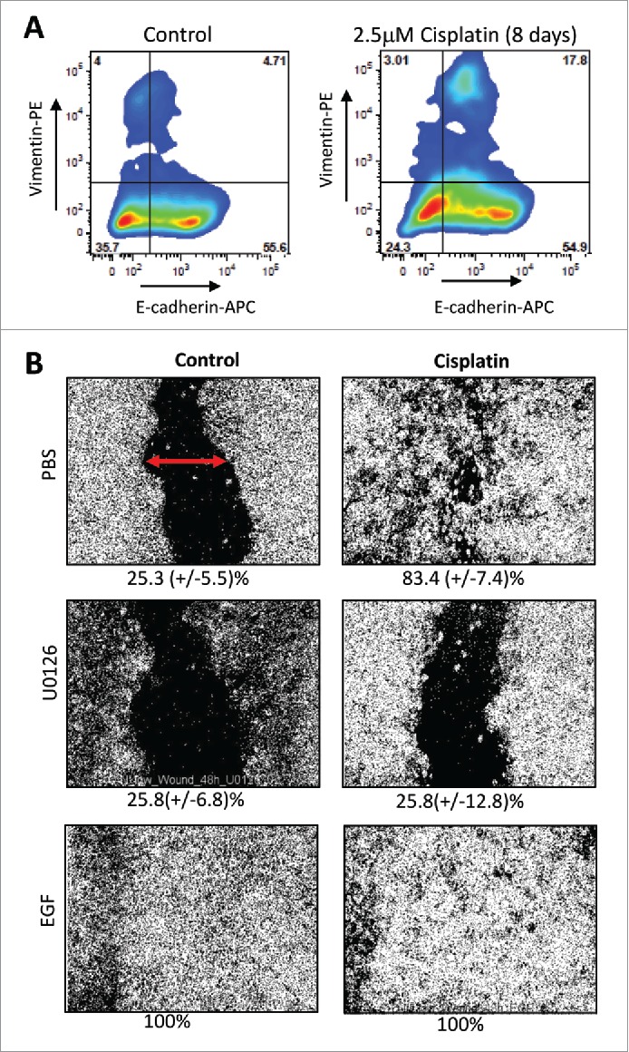
Figure 4.

Effect of cisplatin on epithelial-mesenchymal phenotype in ovarian cancer. (A) Flow cytometry analysis for E-cadherin and Vimentin on untreated ovc316 cells and cells treated with 2.5μM cisplatin for 8 days. Representative studies are shown. (B) Wound healing assay. The migratory ability of ovc316 cells was evaluated by a “wound-healing” assay. A scratch was made with a sterile tip in a confluent layer of cells (marked by red arrow) and “wound” closure was observed after 48h of 2.5μM cisplatin treatment. The MAPK inhibitor U0126 (10 μM), was used to block cell migration. As a positive control, EGF (100 ng/ml), a known MAPK pathway activator was used. Wound closure was measured with ImageJ. Representative images are shown. The percentage of wound closure is shown beneath the images. N = 3.
