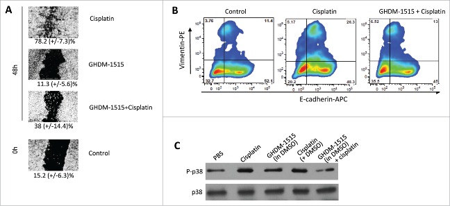
Figure 8.
GHDM-1515 counteracts changes triggered by cisplatin in ovc316 cells. (A) Wound healing assay. Assay conditions were as described for Fig. 3. The concentration of GHDM-1515 was 2 μM. Representative image is shown. The percentage of wound closure is shown beneath the images. N = 3. (B) Flow cytometry analysis for E-cadherin and Vimentin was performed as described in Fig. 3. Cells were treated with cisplatin or cisplatin + GHDM-1515 for 4 days. (C) Activation of p38 MAPK. Ovc316 cells in 12-well plates (0.5 ×10⁶ cells per well in 2ml of medium) were treated with PBS, 2.5μM cisplatin, 2μM GHDM-1515 (1000x stock in DMSO), 2.5μM cisplatin +2μl DMSO, and 2.5μM cisplatin +2μM GHDM-1515. Cells were collected 3 days later and lysates were analyzed by Western blot with antibodies specific to the phosphorylated form of p38 (P-p38) (upper panel). Filters were then stripped and incubated with p38-specific antibodies (lower lane).

